Home
/ Blood Vessels Labeled - Great Vessels Wikipedia - Name the blood vessel labeled 'c'.
Blood Vessels Labeled - Great Vessels Wikipedia - Name the blood vessel labeled 'c'.
Blood Vessels Labeled - Great Vessels Wikipedia - Name the blood vessel labeled 'c'.. Arteries (in red) are the blood vessels that deliver blood to the body. Blood vessels are an integral component of the circulatory system. The function and structure of each segment of the peripheral vascular system vary depending on the organ it supplies. We did not find results for: There are three distinct layers forming the walls of arteries and veins.
Blood vessels differ slightly in location from cat to cat. The iliac, femoral, popliteal and tibial (calf) veins are the deep veins in the legs. Maybe you would like to learn more about one of these?. Navigate to the cardiovascular system area in the following pal 3.0 module: The vessels that carry blood away from the heart are called arteries, and their very small branches are arterioles.
Wire Models from classroom.sdmesa.edu The walls of blood vessels differ depending on the type of vessel. Check spelling or type a new query. Digestive tract of human 12 photos of the digestive tract of human digestive system of human body parts and functions, digestive system of human in tamil, digestive system of human in urdu dailymotion, digestive system of human notes, digestive tract of human, inner body, digestive system of human body parts and. Small vessels that supply blood to outer part of the larger vessels. The vessels that carry blood away from the heart are called arteries, and their very small branches are arterioles. Blood vessel structure and function. The vessels make up two closed systems of tubes that begin and end at the heart.one system, the pulmonary vessels, transports blood from the right ventricle to the lungs and back to the left atrium.the other system, the systemic vessels, carries blood from. Name the blood vessels labeled 'e'.
The vessels make up two closed systems of tubes that begin and end at the heart.one system, the pulmonary vessels, transports blood from the right ventricle to the lungs and back to the left atrium.the other system, the systemic vessels, carries blood from.
Blood vessels consist of arteries, arterioles, capillaries, venules, and veins. Blood vessel labeling 7p image quiz. There are three distinct layers forming the walls of arteries and veins. Structure & function of blood vessels. Blood vessels of the head and neck last updated on fri, 02 jul 2021 | anatomy and physiology blood is supplied to parts within the neck, head and brain through branches of the subclavian and common carotid arteries. Blood cells by descartes 48,739 plays 9p image quiz. Deep veins, located in the center of the leg near the leg bones, are enclosed by muscle. Arteries, veins, and capillaries blood vessels flow blood throughout the body. Blood vessels labeled diagram, blood vessels labeling exercises, cat blood vessels labeled, human anatomy blood vessels, human. The venules and veins returning blood to the heart. Blood pressure is measured as two readings, systolic and diastolic. Blood circulates throughout the body in blood vessels, propelled by the pumping action of the heart. What is the name of blood vessel b?
Blood vessels are the channels or conduits through which blood is distributed to body tissues. Veins (in blue) are the blood vessels that return blood to the heart. Maybe you would like to learn more about one of these?. While most blood vessels are located deep from the surface and are not visible, the superficial veins of the upper limb provide an indication of the extent, prominence, and importance of these structures to the body. The thick outermost layer of a vessel (tunica adventitia or tunica externa ) is made of connective tissue.
4a2a6 15grw 3xjnd8mq3uiet6aug Brains Blood Vessels Labeled Hd Png Download Vhv from www.vhv.rs The iliac, femoral, popliteal and tibial (calf) veins are the deep veins in the legs. The function and structure of each segment of the peripheral vascular system vary depending on the organ it supplies. Spend a few minutes reviewing the name and location of each one, then try testing your knowledge by filling in your own diagram of the respiratory system (unlabeled) using. Name the blood vessel labeled 'b'. This article lists a series of labeled imaging anatomy cases by system and modality. Blood vessels are an integral component of the circulatory system. Check spelling or type a new query. Classification & structure of blood vessels.
The venules and veins returning blood to the heart.
Related posts of the human blood vessels labeled digestive tract of human. Very small branches that collect the blood from the various organs and parts are called venules, and they unite to form veins, which return the blood to the heart. Name the blood vessel labeled 'b'. Name the blood vessels labeled 'e'. Dr calum worsley and assoc prof craig hacking et al. Classification & structure of blood vessels. This layer is lined by endothelium, which is comprised of simple squamous epithelial cells. Maybe you would like to learn more about one of these?. Blood vessels 11p image quiz. Arteries (in red) are the blood vessels that deliver blood to the body. Larger arteries and veins contain small blood vessels within their walls known as the vasa vasorum —literally vessels of the vessel—to provide them with this critical exchange. While most blood vessels are located deep from the surface and are not visible, the superficial veins of the upper limb provide an indication of the extent, prominence, and importance of these structures to the body. Veins return blood back toward the heart.
Arteries and veins are composed of three tissue layers. They are found in bone marrow, the spleen, the liver and the maternal side of circulation within the placenta. Hma practical 3 for monday july 23 and wednesday july 25. B.mention three adaptation of plant to reduce excessive transplration. Digestive tract of human 12 photos of the digestive tract of human digestive system of human body parts and functions, digestive system of human in tamil, digestive system of human in urdu dailymotion, digestive system of human notes, digestive tract of human, inner body, digestive system of human body parts and.
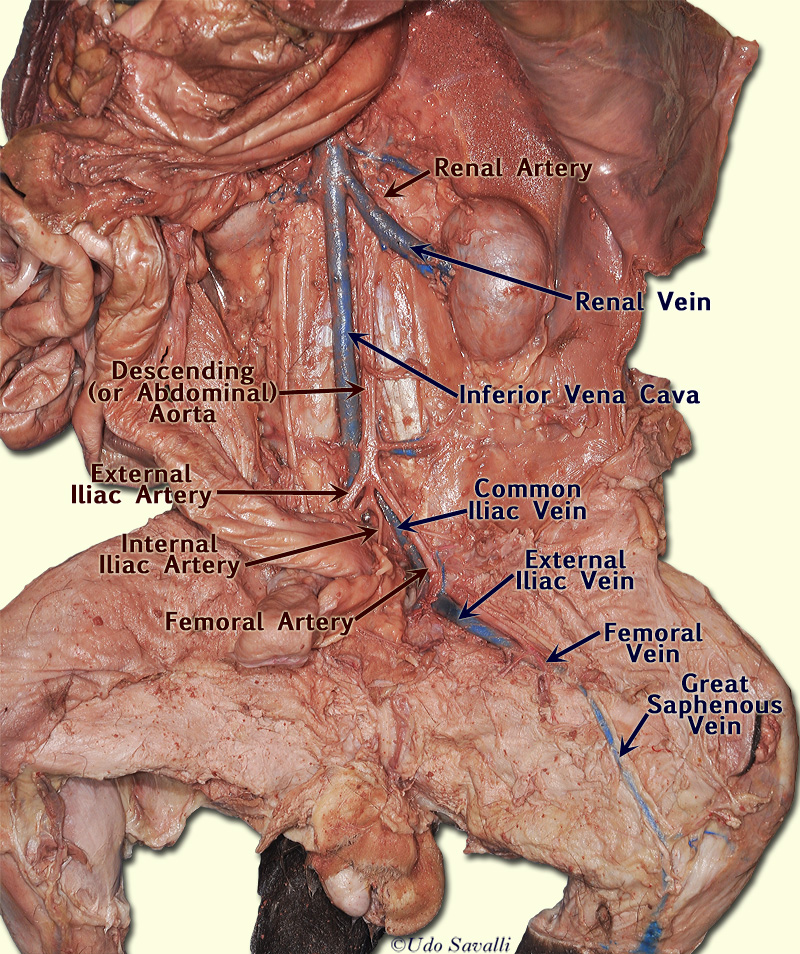
Bio202 Cat Vessels from www.savalli.us The common cartoid artery extends from the brachiocephalic artery. Veins (in blue) are the blood vessels that return blood to the heart. Small vessels that supply blood to outer part of the larger vessels. This article lists a series of labeled imaging anatomy cases by system and modality. Blood pressure is measured as two readings, systolic and diastolic. Found in kidneys, intestines, and endocrine glands. Structure & function of blood vessels. The walls of blood vessels differ depending on the type of vessel.
The venules and veins returning blood to the heart.
•formed where capillaries unite • extremely porous 1) venules: Blood vessels form a continuous path for blood flow that starts and ends at the heart.arteries carry blood away from the heart, regardless of the degree of blood oxygenation.veins carry blood toward the heart. Blood vessels consist of arteries, arterioles, capillaries, venules, and veins. Name the blood vessel labeled 'b'. Introduction to heart and blood vessel disorders in cats cat owners merck veterinary manual : The thick outermost layer of a vessel (tunica adventitia or tunica externa ) is made of connective tissue. Name the blood vessels labeled 'e'. The adventitia or outer layer which provides structural support and shape to the vessel This set is often in folders with. Arteries (in red) are the blood vessels that deliver blood to the body. Check spelling or type a new query. Veins return blood back toward the heart. Blood vessels 11p image quiz.

